About this episode
Get the Book now from Amazon:
https://www.amazon.com/dp/0323624367?lv=shuf&channelId=500&plpRedirect=mhFallback&linkCode=ll2&tag=cvthunderx-20&linkId=779be838f7807ad3e9f2b1cf1d8aebd4&language=en_US&gaOptInStatus=true&ref_=as_li_ss_tl
You can listen and download our episodes for free on more than 10 different platforms:
https://linktr.ee/dentistry_made_simple
Produced by:
https://www.podcaistudio.com/
Get every episode summarized
Each time Dentistry Made Simple publishes, we email you a written briefing from the transcript — the topics, who appeared, and any specific claims, with the ad reads skipped.
Email me new episodesFree for 3 shows. No card needed.
Hosts & guests
Transcript ready
756 searchable segments. Every word is indexed and playable.
Full transcript
Dentistry Made Simple — Endodontics: Principles and Practice. Machine-transcribed; use the interactive transcript above to jump the player to any line.
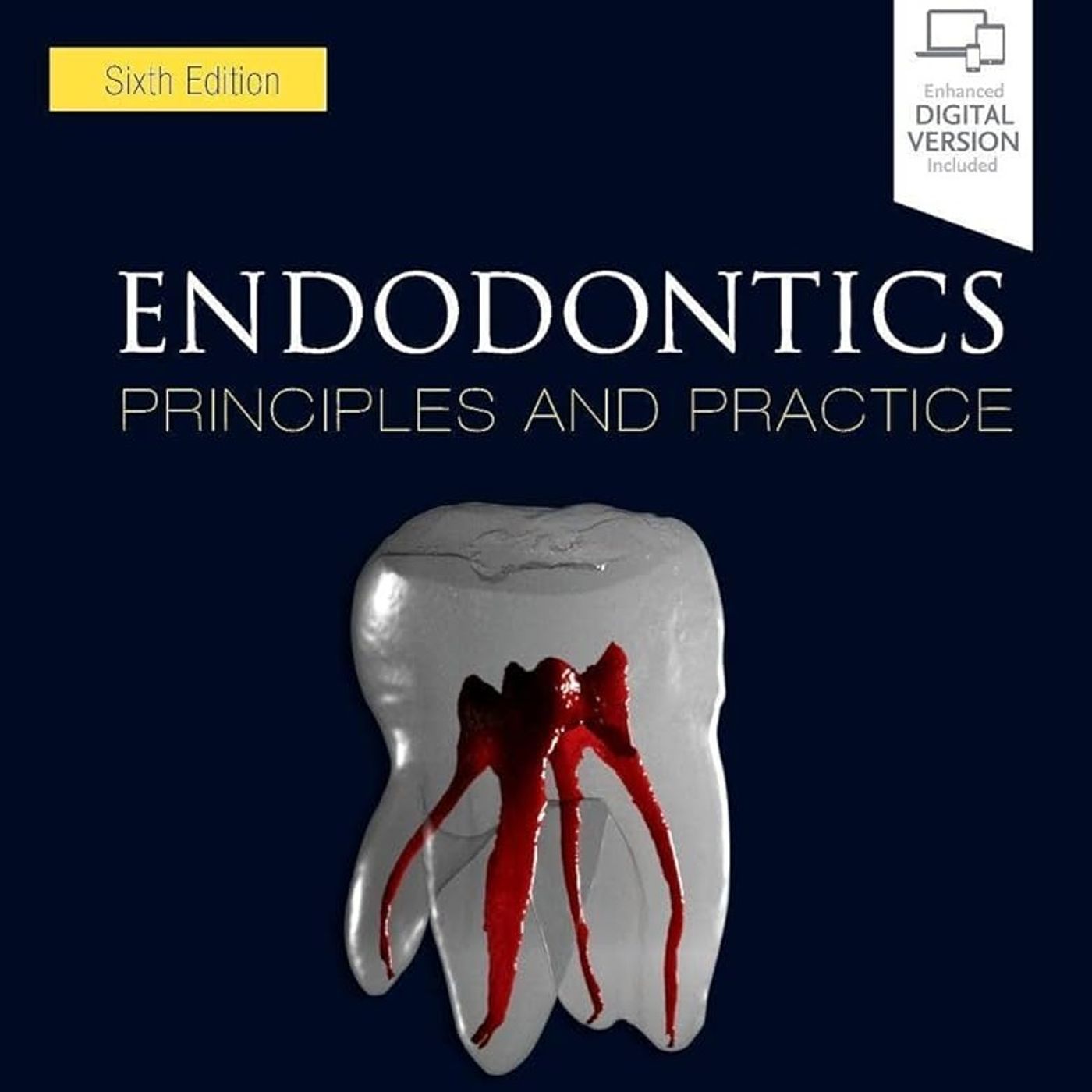
So imagine like a microscopic fortress, it's built entirely out of Denton, which is one of the hardest substances in the human body, completely rigid. Right, totally sealed off. Exactly, mostly sealed off from the outside world. But inside that fortress is this incredibly delicate, highly sensitive living ecosystem. And for most of a patient's life, that fortress does its job perfectly. But the second those walls are breached, whether by decay or physical trauma, that beautiful ecosystem turns into an absolute pressure cooker. It really does. It's essentially the ultimate biological locked room mystery. You have this vital tissue trapped in a rigid chamber, right? And when inflammation hits, the normal physiological response, which is swelling, just, well, it has absolutely nowhere to go. And navigating that exact scenario is, you know, what we are unpacking today. Welcome to the deep dive. We are pulling some really critical insights from the sixth edition of Endodontics Principles in Practice. That's by Doctors, Toro Binajad, Fuad, and Shabahang.
Yeah, it's a phenomenal textbook. It really is. And we're looking closely at the overarching philosophy of this text, which is that the primary goal of Endodontics is to relieve pain and preserve the natural dentition. It's this constant balancing act between the art of clinical execution and the actual science of biological evidence. Which makes it just a crucial resource for anyone holding a handpiece, you know? I mean, you might think of Endodontics as the strict domain of the specialist. But the clinical data tells a totally different story. Right, general practitioners do most of them. Yeah, they actually perform about 75% of all root canal treatments. So having a specialized level understanding of this material isn't just like a bonus for a general dentist, it is a strict clinical requirement for daily practice. Exactly. So for you listening, whether you're a student or a practicing clinician, we are going to trace the entire life cycle of a case. We'll start inside that microscopic fortress of the pulp, move through the complexities of diagnosis, explore this massive biomechanical debate
that's currently shifting how we shape teeth, and finally look at how modern surgery dictates whether we actually succeeded or not. Sounds like a solid roadmap. Yeah, so let's start at the very beginning with the biology. When you look at the dental pulp, it's a highly specialized environment, right? It originates embryologically from neural crest cells. It does, yeah. And if you look at it histologically, it is a specialized connective tissue. It contains fibroblasts, undifferentiated mesenchymal cells, and a really dense structural matrix. Made mostly of collagen, right? Yes, specifically types I and II, along with proteoglycans, glycoproteins, and crucially, an incredibly high water content. Okay, so wait, if the matrix is mostly water and collagen, how does it handle the onset of inflammation? Because normally, you know, when tissue gets infected, it swells up to allow immune cells to flood the area. Right, and that right there is the core physiological problem of the pulp. Because it's encased in rigid dentin, it literally can't expand. Wow, okay. So when bacteria cause information,
the localized tissue pressure rises so rapidly, and this increased pressure can actually collapse the thin wall venules inside the pulp. Which cuts off the blood flow. Exactly, the blood can't escape, the tissue becomes hypoxic, and you get this localized necrosis that just start spreading throughout the chamber. But the pulp isn't just like passively waiting to die, is it? Because the text points out that the cells standing on the front lines, the odontoblasts, they aren't just laying down dentin, they are actively fighting off invaders. Oh, yeah. That's actually one of the most fascinating aspects of pulp biology. The odontoblasts act as the pulp's very first line of innate immune defense. They form this tightly packed, palisating layer right up against the dentin, and they express toll-like receptors. So how do those receptors actually function in that specific environment? Well, think of toll-like receptors as biological trip wires. They recognize pathogen-associated molecular patterns. Which are basically the structural signatures of bacteria.
Spot on. So when decay nears the pulp, and these receptors detect those bacterial signatures, the odontoblasts immediately release cytokines and defensins. Oh, wow. Yeah, they essentially sound the biochemical alarm and start neutralizing pathogens before the bacteria even physically breach the pulp chamber. That is wild, and that actually brings us to the alarm system itself, because the nerve fibers transmitting this information are critical for you to understand when a patient is actually sitting in your chair. Absolutely. You have two main types of sensory nerves, right? The Adelta fibers and the C fibers. And reading through this, it kind of reminds me of a two-tier home security system. I like that analogy. Yeah. Adelta fibers are located out in the periphery, right? Extending into the dentinol tubules. And they are myelinated, which means they transmit signals incredibly fast. They are like the glass break sensors on your windows. Perfect analogy, yeah. The myelons sheath allows for rapid saltatory conduction. So when you place a piece of ice on a vital tooth,
it's those Adelta fibers firing that sharp, immediate, shocking pain. Right, the stuff that makes the patient jump. But then you have the C fibers, which are more like the slow burn interior motion detectors. They are unmyelinated and located much deeper in the central pulp. Right, they don't give you that fast shock. Exactly. They give you that deep dull, agonizing, throbbing pain. The kind that keeps the patient awake all night long. Which is the worst. And understanding which fiber is firing really dictates how you interpret your patient's symptoms. Now, if we catch an injury early, say, I don't know, you have a traumatic exposure of a vital bleeding pulp in a young patient. We don't necessarily have to jump straight to complete extrapatient and a full root canal. Right, the text highlights vital pulp therapy for that, specifically the partial polypotomy. So let's talk about the biological mechanism behind that protocol. Because if I'm looking at a traumatic exposure, my initial instinct might just be to clean the whole chamber out so I don't trap any bacteria.
How do you manage a partial polypotomy without just accidentally sealing in an infection? Well, the primary goal here is mature genesis. We want to preserve healthy tissue to allow the root to actually finish developing. So you start by amputating just the superficial inflamed pulp. Using a specific burr. Yeah, what a cool diamond burr at high speed. Because you want clean cutting, no tearing, you have to minimize cellular damage. And then you achieve human statesis and disinfect the area by applying sodium hypochloric. Okay, and then comes the chemical conditioning, which the text emphasizes heavily. You apply 17% EDTA. But why is that specific chemical so necessary in this step? It's all about the smear layer. I mean, when you cut dent in with a burr, you leave behind this microscopic sludge of pulverized hydroxyapatite, collagen, and potentially bacteria, right? Right, which just plugs up all the diktinal tubules. Exactly. And EDTA is a chelating agent. So it strips away the calcium, dissolves that smear layer entirely and unplugs the tubules. So you are basically opening up the microscopic pores
of the tooth. Yeah, exactly. Because once you dry it, you cap that vital pulp with a bioactive tricalcium silicate material. And if the tubules are open, this biosuramic can interface perfectly with a dentin. Oh, that makes total sense. Plus, as the material sets, it releases calcium hydroxide, which is highly alkaline. So this not only creates an antibacterial environment, but it actively recruits undifferentiated stem cells to migrate to the site. Wait, really? It recruits stem cells. Yeah, they migrate over and form a hard tissue bridge, essentially permanently sealing the pulp back off on its own. That is just incredible tissue engineering. But what about trauma that doesn't expose the pulp at all? Say it's fresh and gets hit in the mouth, suffering lexation injury. And over a few years, the tooth just turns completely yellow. You take an x-ray, and the canal is just completely obliterated. Hulsific metamorphosis. Right, very common. Again, my instinct as a clinician would be to drill in and try to find that canal before it disappears entirely and becomes just impossible to treat. It's a very common instinct, for sure.
But the text establishes a hardened fast rule here. Calcific metamorphosis on its own does not indicate a need for root canal treatment. Really? You just leave it. You have to remember the biology, you know? The pulp is vascularized. It is laying down tertiary dentin rapidly in response to the trauma to protect itself. It's a defense mechanism, not a disease state. Oh, OK. So unless you see clinical or radiographic signs of necrosis-like, say, an apical radioleucency, you just leave it alone. Interveating too early just invites Iatrogenic errors, like, you know, preferring the root, trying to find a tiny canal that is actually functionally healthy. Which perfectly illustrates why diagnosis is so difficult, and honestly, why guessing is just so dangerous. But identifying whether a tooth is vital or calcified is really only half the battle. If a patient is in absolute agony, but the visual exam looks completely normal, how do we actually find where the pathology is hiding? Well, diagnostics is where the true science of endodontics really shines. I mean, you simply cannot treat what you have not
accurately diagnosed. Let's talk about sensibility testing them, because the terminology here really matters. If I want to know the status of a pulp, the text points to cold testing, specifically using a refrigerant like endo-ice as being the most accurate test. It is the most accurate, yeah. But we have to be really precise clinically here. Cold testing or using an electric pulp test in EPT, those are sensibility tests. Meaning they don't test blood flow. Exactly. They only test whether the nerve fibers, those A delta and C fibers we talked about, are capable of firing an action potential. They do not directly test true pulp vitality. Because true vitality refers to vascular blood flow. Right. Which you would need, like a laser Doppler to actually measure. However, in a normal clinical setting, nerve response is really our best proxy. And the text notes that combining cold testing with EPT yields the absolute highest diagnostic accuracy. Because you are testing the neural response through two completely different mechanisms. But the real diagnostic revolution isn't the endo-ice.
It's the imaging. The shift from standard 2D periapical x-rays to 3D cone beam computer tomography or CBCT is just changing everything. Oh, completely. The clinical stats in the text are actually wild. When clinicians evaluate a case using a CBCT scan, it alters their clinical treatment plan in 27.3% of general cases. And in high difficulty cases, it changes the plan over 50% of the time. It really is a massive paradigm shift in how we see pathology. Because a big problem with 2D imaging is superimposition. Right. Everything is just flattened. Yeah. For you to see a periapical lesion on a standard radiograph, that infection has to literally erode all the way through the inner cortical plate of the jawbone. If the lesion is just sitting inside the cancelist bone, the dense cortical bone on the x-ray simply masks it. It just hides it completely. Plus, you have all these anatomical structures like the zygomatic process obscuring the maxillary molars. So a patient could have this massive infection, but on a 2D film, it looks like perfectly healthy bone.
Precisely. Yeah. But CBCT gives you slices. You essentially remove all that anatomical noise. You can see the true 3D extent of external cervical root resorption. You can find hidden MB2 canals that just overlap on 2D films. And you can assess the exact dimensions of a bone lesion in all three planes of space. Exactly. OK. But here's where I have to push back a bit. If we know that a 2D film misses lesions in the cancelist bone, and we know that looking at a 3D scan changes our treatment plan half the time in complex cases, shouldn't we just mandate a CBCT for every single endodontic patient who walks through the door? I mean, why ever guess with 2D if 3D is that much better? It's a very logical conclusion. I totally get that. But it ignores a really fundamental tenant of radiology that governs our standard of care, which is the LRA principle. As low as reasonably achievable. Right. A CBCT, even with a really small field of view, delivers more ionizing radiation to the patient than a standard digital pariapical radiograph. So the standard of care, supported by the AIE,
dictates selective use. So if you have a pretty straightforward diagnosis on, say, a maxillary central incisor with a massive obvious lesion, a 2D radiograph gives you all the information you need to proceed safely. Exactly. You reserve the CBCT for cases where the 2D information is contradictory to the patient's symptoms. Or for complex morphology, resorbed of defects, retreatment of failing cases, or if you suspect a vertical root fracture. Got it. You only expose the patient to that extra radiation when the 3D data will definitively change or guide your management of the case. Of bottom. And we can't forget that diagnosing an endodontic case isn't just about staring at teeth or clicking through imaging software. The text heavily emphasizes the physical, extra-all examination, especially during emergencies. Like before you even look inside the mouth, you have to look at the patient's face. Absolutely. Because you are evaluating for systemic infectious spread, are there sub-menibular lymph nodes palpable? Do they have restricted mouth opening toismus due to muscle spasms from the infection?
Or is there obvious facial cellulitis? Right. An endodontic infection isn't always safely localized to the alveolar bone. If it breaches the cortical plate, it can spread right into the facial spaces of the head and neck. And that can rapidly become a life-threatening airway issue. Which is terrifying. It is. Identifying the systemic involvement dictates whether you can just simply open the tooth or if you need to prescribe aggressive antibiotics and essentially manage a severe medical emergency. OK. So assuming we have completed all our detective work safely, we have a definitive localized diagnosis of irreversible pulpitus or necrosis. Now we actually move to the chair. And I want to start with a bit of a reality check regarding the standard of care. This is such a vital medical legal and ethical point. We established earlier that general practitioners do the vast majority of root canals. Well, the text is unindiguous here. The standard of care is identical, regardless of who holds the handpiece. Wow. Yeah. A general dentist is held to the exact same standard of clinical excellence and biological outcome
as a board certified endodoncist. Which really highlights why the AIE case difficulty assessment form is such a critical tool. It allows you to objectively look at a case, the root curvature, the degree of calcification, maybe the patient's limited mouth opening, and decide, do I have the equipment and the psychomutter skills to treat this to the specialist standard or do I really need to refer this out? Right. It takes the ego out of it. Exactly. But how do we actually train our hands to meet that high standard? Honestly, it's one of the hardest transitions in dental education. You can memorize the biology all day long. You can understand the 3D anatomy, but that doesn't automatically teach your hands what your mind knows. Right. The tactile feel. Yeah. You are operating in a microscopic space, often relying entirely on tactile feedback and a tiny mirror. If any of these all your movements are reversed, that's why preclinical practice on extracted teeth and specialized 3D printed models is just non-negotiable. You literally have to put the reps in. You do. You have to physically learn that subtle, drop feeling when a file actually enters the pulp chamber
or the specific gritty resistance of a calcified canal. And speaking of entering the pulp chamber, this brings us to one of the most intense biomechanical debates in modern endodontics right now, the access cavity. Oh, yes. The access debate. It's everywhere. It is. Historically, the absolute gold standard was straight line access. You remove enough coronal tooth structure so that your files can drop straight down into the apical third without bending it all. Which makes sense mechanically. It does. It drastically reduces the cyclic fatigue on your rotary instruments, meaning you are much, much less likely to separate a file in the canal. And it makes mechanical debridement highly efficient. But the downside is massive, right? Because to get that straight line path, you are aggressively removing peri-servical dentin. And we now know that peri-servical dentin, which is the dentin roughly four millimeters above and below the crestal bone, is the crucial structural backbone of the tooth. It really is. It acts like the metal hoops on a wooden barrel, right?
Distributing the stress of chewing. So if you hollow it out, sure, you get a perfectly clean root canal, but the tooth structurally fails and snaps in half two years later. Exactly. So recently, the pendulum has swung heavily toward contracted endodontic cavities. Basically, making the access hole is absolutely tiny is possible. The goal is to preserve that peri-servical dentin, to maximize the fracture resistance and long-term survivability of the tooth. But wait, if I make the access cavity so incredibly small that I can't even easily see the pullporns, aren't I severely compromising my ability to actually clean the tooth? If I leave necrotic tissue and biofilm behind, just because I simply couldn't reach it, the tooth might be structurally strong, but the root canal will fail biologically anyway. And you have to start to take you away to the exact tension driving this massive debate in the literature. It is a huge trade-off. Preserving tooth structure is incredibly important, obviously. But the primary biological goal of root canal therapy is the elimination of microbes. Right. If your contracted access prevents adequate disinfection,
the treatment is a failure. Full stop. The text suggests a balanced approach. You would have served dentin wherever possible, but never, ever at the expense of adequate cleaning, shaping, and visualization. OK, so let's say we've compromised perfectly. We've cleaned the canal, removed the biofilm, and shaped it to a nice continuous taper. Now we have to fill it. And the specific word the text uses for the goal of obteration is fascinating to me. They call it intuming. It's a very intentional word choice, for sure. Because despite our absolute best efforts with ultrasonic irrigation and gallons of sodium hypochloride, it's biologically impossible to sterilize a root canal system completely. There's always something left. Always. Bacteria will always survive deep inside the microscopic tetanutubules or hiding complex lateral canals. So the goal of obteration isn't true sterility. It's intumant. We're basically trapping them. Exactly. We want to fill that 3D space so completely that we entune the remaining bacteria in a fluid tight seal of getter perch and sealer.
We cut them off from tissue fluids and nutrients, so they simply cannot proliferate. And the technology for doing this has evolved pretty significantly. The text highlights carrier-based obteration. How does that achieve a better seal than just packing coal cones of getter perch into the canal? Well, carrier-based systems utilize a core carrier. And modern versions use a rigid cross-linked gutter perch a core. It gets heated up in a specialized oven, and then you slowly insert it into the shaped canal. OK, so the core itself is pushing the material. Yes. Because the core is cross-linked, it remains rigid enough to push the surrounding flowable heated gutter perch a laterally. It acts exactly like a hydraulic piston, driving the material deep into all the complex fins, webs, and lateral anatomy of the canal system. It creates a far superior three-dimensional entombment. But here's where we have to look at the harsh reality of clinical practice. Because as amazing as our techniques are, the text explicitly states that some bacterial species can actually survive entombment.
They absolutely can. Sometimes, despite a perfectly tapered, beautifully filled x-ray, the apical infection just persists. And when non-surgical retreatment isn't viable, or maybe has already failed, we have to approach the infection from the opposite direction. We essentially have to go through the bone. Right. Which brings us to epical microsurgery. And the terminology here is really important to note. We rarely use the term apicalectomy anymore. The text refers to it as micro-apical surgery, because the precision of the procedure has just evolved dramatically. It's no longer just blindly hacking off the root tip. Far from it, historically, success rates were honestly unpredictable. But today, with the use of operating microscopes and highly specialized instruments, success rates are routinely in the 90% range. Let's look at that surgical cassette for a minute, because the instruments are incredibly specialized to prevent tissue trauma. We aren't just taking a standard scalpel and roughly reflecting the tissue. No. Standard surgical blades can be way too aggressive for the delicate papillia of the jingiva.
That leads to severe recession and scarring. So we use microblades for precise, minimally invasive incisions. And to actually reflect the tissue. To reflect the periodicity of off the bone without tearing it, we use specialized mole curates. They're designed to gently release the tissue rather than aggressively scraping it. But the tool that really stood out to me in the text is the drill used to actually remove the bone and access the root tip. The impact air surgical handpiece. It looks completely different from a regular drill. It has this distinct 45 degree angle. Yeah, the 45 degree angle is crucial because it provides the clinician with direct line of sight visibility into the surgical crypt. With a standard handpiece, the bulky head of the drill completely blocks your view under the microscope. Oh, that makes sense. But it's not just about visibility, right? The text specifies it is uniquely designed not to release exhaust air into the surgical site. Why is that mechanical detail? It's so critical. Because of a really severe complication called surgical emphysema.
OK, what is that? Well, if a standard high speed handpiece exhausts highly pressurized air into an open bony wound, that air can actually be forced deep into the fascial planes of the face and neck. Oh, wow. It causes immediate massive swelling. And you can even track all the way down into the media's genome in the chest, creating an absolute life-threatening emergency. That is terrifying. It is. So the impact air handpiece is engineered to direct the exhaust air backward, out the rear of the hand piece, completely eliminating that risk. OK, so we've safely made a window in the bone, no air trapped, and we've identified the infected root tip. We resect three millimeters off the end of the root, right? Because anatomically, that's where the vast majority of apical deltas and complex lateral canals hide all the bacteria. Correct. Then we have to seal the root from the bottom up. Right. This is the retro-preparation phase. We use specialized diamond-coded ultrasonic tips that are angled to perfectly access the long access of the root tip. And they come varying lengths, three millimeters up to six
millimeters, or even nine millimeters. Wait, nine millimeters. You're practically cleaning out half the root system from the bottom at that point. You are. And that is actually necessary in cases where you have, say, a massive, irretrievable metal post in the canal, or highly complex anatomy that just couldn't be negotiated non-surgically from the top down. You clean that space ultrasonically, then seal it with a highly biocompatible material typically MTA, or a biosuramic putty to create a permanent apical plug. So taking a step back from all of this clinical detail, how do we actually know of all this incredible effort, the tricky diagnostics, the biomechanical shaping, the delicate microsurgery? How do we know if it actually worked? We really need to talk about how we define success in modern endodontics. Yeah. And this has changed a lot. The classic definition of success has almost always been purely radiographic. Right, looking at the x-ray. Yeah. If a patient presented with an apical period tightest lesion like a dark shadow on their x-ray success, basically meant the patient was asymptomatic. And over time, that dark shadow
filled back in with healthy, transbacular bone, radiographic healing. But the text introduces a massive shift in how we actually evaluate these outcomes today. It's a modern, deeply patient-centered metric. It's called functional retention. And this is such a crucial evolution in our thinking. Because recent clinical data shows that when root canal treated teeth are eventually extracted, it is very, very rarely because the endodontic infection returned. Really? Yeah. It is overwhelmingly because the tooth suffered a catastrophic vertical root fracture. Let me try to synthesize this with an analogy to only if this tracks. Imagine the tooth is a concrete bridge. OK. The bacterial infection is like rust eating away at the steel rebar inside the pillars of the bridge. The classic radiographic definition of success basically just asked, did we eliminate all the rust? But the functional retention definition asks, OK. We got rid of the rust, but did we hollow out the pillars so aggressively during our cleaning that the bridge just collapses the very next time a heavy truck drives over it?
That is a phenomenal way to conceptualize it. Yes. Functional retention means the tooth is successfully bearing the occlusal loads of chewing. It remains completely asymptomatic. And it is surviving as a functional unit in the mouth long term. It has to actually work as a tooth. Exactly. It doesn't mean we just ignore radiographic healing. We absolutely still want to see bone regeneration down there. But it means we recognize that a perfectly beautiful white line of gutted perch on an x-ray means absolutely nothing to the patient if the tooth fractures in half a week later. It forces us to permanently balance the biology of disinfection with the real world biomechanics of survival. Exactly. Which ties perfectly back into our access debate from earlier. We have to respect and preserve that pericervical dent and do you sure the bridge can still hold traffic? Makes total sense. All right. Before we wrap up this deep dive into the text, it's time for a quick review exercise for you listening. We want to make sure these clinical concepts actually stick. So I want you to mentally recall two specific mechanisms
we covered today. First, define the clinical difference between Adelta fibers and C fibers in the dental pulp. Think back to our home security analogy. Second, what is this specific chemical irrigant and its exact concentration used to remove the smear layer during a partial pulpotomy? And why does it allow the biosuramic to work? OK. So if you recall that Adelta fibers are myelinated and trigger that fast, sharp pain out in the periphery, while C fibers are unmilinated and produce that dull, throbbing pain centrally, you are spot on. And for the chemical. That would be 17% EDTA, which chalates calcium to unplug the dental tubules. Perfect. So we started this exploration looking at the pulp as an impenetrable fortress, a rigid chamber that protects but also traps its vital tissue. We explored the biology of how to navigate it, how to shape it without destroying its structural integrity, and how to surgically intervene when entombment inevitably fails. But I want to leave you with a final thought to mull over in your next clinical session or study group. Let's hear it.
If the ultimate goal is permanently shifting, if functional retention and a tooth's biomechanical susceptibility to fracture are now universally recognized as being just as important as radiographic healing, how is this going to shift the future design of endodontic instruments? I mean, if we simply cannot make the access larger without fatally weakening the tooth, will we eventually see the rise of highly flexible, shape, memory, smart files? Or perhaps like completely touchless, acoustic cleaning technologies that can navigate and disinfect the fortress without ever needing to tear down its walls at all? That is the big question. Keep asking those questions as you practice. Thanks for joining us on this deep dive, and we'll catch you next time.
More episodes
More from Dentistry Made Simple

Endodontic Radiology
Dentistry Made Simple

Emergency Medicine Oral Board Review: Pearls of Wisdom
Dentistry Made Simple

Facial Aesthetics: Concepts and Clinical Diagnosis
Dentistry Made Simple

Essentials of Pharmacology for Dentistry
Dentistry Made Simple