Loading...
About this Episode
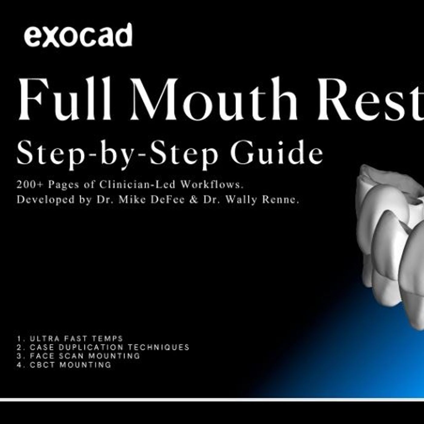
A comprehensive guide to modern full mouth restorative dentistry using advanced digital workflows. Developed by Dr. Wally Renne and Dr. Mike DeFee, the text details how to utilize CAD software and 3D printing to create precise pre-surgical waxups and life-like prototypes. The authors emphasize that moving away from traditional methods toward digital design increases both predictability and clinical efficiency. Key technical procedures mentioned include face scan mounting, CBCT integration, and the development of ultra-fast temporary restorations. Ultimately, the guide serves as an instructional resource for clinicians to blend technological innovation with artistic dental care.
You can listen and download our episodes for free on more than 10 different platforms:
https://linktr.ee/dentistry_made_simple
Produced by:
https://www.podcaistudio.com/
You can listen and download our episodes for free on more than 10 different platforms:
https://linktr.ee/dentistry_made_simple
Produced by:
https://www.podcaistudio.com/
Hosts & Guests
Transcript
Are you really buying a car online on auto trader right now?
Really?
At a playground?
Yeah, really.
Look at these listings from dealers.
Wow.
Your search can really get that specific.
Really?
And you just put in your info and boom.
Cars in your budget.
Mom needs a second, honey.
You can really have it delivered?
Really.
Or I can pick it up with the dealership.
One sec, sweetie.
Mommy's buying a car.
Mommy's working.
I think kid is walking up the slide.
Kyle, again, really?
Auto trader.
Buy your car online.
Mommy's buying a car.
Mommy's working.
I think kid is walking up the slide.
Kyle, again, really?
Auto trader.
Really?
At a playground?
Yeah, really.
Look at these listings from dealers.
Wow.
Your search can really get that specific.
Really?
And you just put in your info and boom.
Cars in your budget.
Mom!
Mom needs a second, honey.
You can really have it delivered?
Really?
Or I can pick it up with the dealership.
Mom!
One sec, sweetie.
Mommy's buying a car.
Mommy's working.
I think kid is walking up the slide.
Kyle, again, really?
Auto trader.
Auto trader.
Buy your car online.
Really?
Welcome to The Deep Dive.
Today, we are looking at something that is quietly revolutionizing healthcare.
And it's actually happening right inside our mounts.
Yeah.
It's a massive shift.
It really is.
We are getting into a fascinating 2024 clinical manual.
It's called DSD Full Mouth.
Right, authored by Dr. Wally Ran and Dr. Mike DeFey.
Exactly.
From the M.O.D. Institute, which is this advanced dental training center out of Charleston,
South Carolina.
And the mission of our deep dive today is to unpack these massive clinical workflows
they have put together.
I mean, we are talking hundreds of pages of highly detailed step-by-step protocols.
And the core of it is understanding how software and facial scanning and 3D printing are completely
replacing old-school dental techniques.
Yeah, whether you are the one holding the drill or you're the one sitting in the chair,
this shift changes the stakes entirely for predictability and aesthetics in dentistry.
Totally.
And we're going to contact this.
Yeah.
Because for decades, dentistry has been stuck in a very specific analog rut.
If you have ever had that cold, gag-inducing tray of putty shoved into your mouth to take
a mold of your teeth.
Oh, yeah.
The physical impression.
You know the trauma.
It is a right-of-passage we would all rather skip, frankly.
And the issue isn't just that it is uncomfortable for the patient, it's that the entire foundation
of a really complex medical procedure is being built on a material that can warp or shrink
or pull.
Right.
If it's a lock-sane or alginate, they have dimensional instability.
Exactly.
Every single physical translation introduces a new margin for deviation.
And from that, putty dentists historically used to make analog temporaries out of materials
like bysacral, which is basically a standard acrylic resin.
I have always wondered how accurate that could possibly be.
You were taking a physical mold, pouring a stone model into it, manually building up
this resin, and just hoping the physical translation doesn't ruin the fit.
It is incredibly prone to human error.
You are hoping the impression material didn't distort when you pulled it out of the patient's
mouth.
You're hoping the dental stone didn't expand too much when it's set.
Right.
The thermal expansion.
Exactly.
And then you are hoping the bysacral resin cures perfectly without shrinking.
What this manual from the MOD Institute introduces is a complete departure from all of that.
It brings us into the era of advanced computer-aided design or CAD.
So dentists are now empowered to craft highly customized, incredibly precise digital
blueprints, well before they ever pick up a drill to prepare it to.
They are.
And it fundamentally changes the workflow.
It feels like we are finally treating the mouth like a high stakes construction project.
If you are building a skyscraper, you don't just start pouring concrete on day one and
cross your fingers that the elevator shafts line up on the 50th floor.
Right.
You would have a comprehensive 3D digital model.
Exactly.
The physics, the load bearing walls, the aesthetics, that is exactly what is happening here.
The clinician can modify and perfect the restorative plan virtually.
By working out all the anatomical kinks on a computer's green first, you are bringing
high predictability to what is frankly a very complex reconstruction of the human jaw.
The architectural analogy works perfectly here.
And the implications for the patient are profound.
Because once you have that perfect digital blueprint, you do not go back to the old analog
hoodie and acrylic to make the temporary teeth.
Right.
The manual highlights that these digital designs are translated directly into what they call
dynamic life like prototype temporaries using high resolution 3D printing.
So instead of a dentist manually sculpting a temporary tooth in your mouth while you
hold your jaw open, they just hit print on a machine down the hall.
Essentially yes.
The clinician 3D prints a physical preview of the final digital result.
Wow.
It completely streamlines the clinical process.
The patient gets a highly accurate test drive of their new smile.
They can go home, eat with it, speak with it, see how it looks in the mirror.
And the clinician knows exactly how the final ceramic restorations will function because
the printed prototype is an exact replica of the digital blueprint.
Let's talk about the software that makes this possible.
Because the manual dives really deep into a CAD program called exocad.
There is a specific phase called the pre-preparation mockup.
This is step one of the major workflow.
Right.
The diagnostic phase.
The authors are adamant that every large full mouth case needs a diagnostic mockup designed
before the patient even shows up for prep day.
A big part of this involves establishing a new vertical dimension.
The vertical dimension of occlusion or video.
Yes.
For anyone unfamiliar, the vertical dimension of occlusion is just the vertical distance
between your upper and lower jaws when your teeth are biting together.
If someone has severely worn down teeth, their face actually collapses slightly, they lose
that vertical height.
But the manual also insists on extensive photography at this stage, which implies we are doing
more than just fixing the bite mechanics right.
We are absolutely doing more than mechanics.
Reestablishing that vertical dimension gives you the foundational architecture for the
entire case.
But the photos provide the necessary facial context.
Because you aren't just designing teeth and avoid.
Right.
You are not floating in empty space on a monitor.
You are designing teeth to fit a specific, moving, expressive human face.
The lips, the smile line, the facial symmetry, all of that dictates where the teeth should
go.
Now here is where I get tripped up when reading through the software workflow.
Specifically around step 15 of the complete mockup wax up phase.
The manual points out a massive trap in the exocad software.
What's fascinating here is the absolute specificity of the software guardrails that authors
emphasize.
It's basically screams at the user.
It says do not hit the button that says adapt design to Jaws Gan hard cut.
And absolutely do not hit the feature to cut all intersections.
But wait, that feels totally backward.
If I'm designing something digitally and I see overlapping digital lines or meshes colliding,
my first instinct as a user is to hit the cleanup button or the auto adapt button.
Why are the authors telling us to leave the mess?
Because it is the equivalent of letting your smartphones auto correct completely rewrite
a nuance sentence you just typed.
It might look a grammatically cleaner to the machine, but it destroys the meaning.
That makes sense.
If you let the software auto adapt or cut all intersections at this diagnostic stage, the
algorithm is going to forcefully alter your carefully planned idealized digital wax up so
that it fits the patient's existing flawed tooth structure.
Because the patient hasn't actually had their teeth prepared or drilled down yet.
Exactly.
So digital teeth are naturally going to intersect with the scans of their current bulkier crooked
teeth.
Precisely.
If you hit that cut button, the software basically subtracts the existing tooth geometry
from your ideal design.
It chops away your ideal shape.
You lose the perfect geometry you just spent hours creating.
You end up with these hollowed out deformed digital shells instead of perfect teeth.
Yes.
It demonstrates how precise the digital workflow must be.
You have to operate within very specific guardrails and understand how the software thinks.
Otherwise you will generate an error that compromises the entire clinical outcome.
That brings up another wild software quirk, the manual highlights for prep day.
This is detailed in steps 16 through 20 and then again around step 107.
The emerging phase.
Right.
The patient is in the chair.
The dentist is finished preparing the physical teeth and they take a new digital scan of
those preps.
Now they need to merge this brand new prep scan with the perfect digital blueprint they
designed days ago.
Which is a critical moment.
But when you import the scan of the prepared teeth, the software throws a pop-up window
at you asking about the coordinate system, specifically if the mesh is relative to the
scan data coordinate system.
And the software practically begs you to let it auto align everything right away.
But the authors are adamant.
You ignore that prompt.
You tell the software, no, if you do that, your 3D models are just floating in digital
space totally disconnected.
I feel like that would induce sheer panic for a clinician.
It would if you don't understand the sequence.
The instinct is to manually drag the models together or force an alignment immediately so
the screen looks organized.
But the manual is teaching restraint.
Because you're dealing with complex data meshes.
Right.
If you force an alignment prematurely or let the software guess based on the wrong coordinate
data, you introduce microscopic misalignments that will ruin the fit of the final teeth.
So you just leave them floating unaligned and click back to the software's main wizard
menu.
Yes.
You trust the software's automated internal processes.
By returning to the wizard, you trigger the proper structured alignment sequence that
the Execad software was built to execute.
And that leads to this visualization tool, the manual describes, which honestly sounds
like a feature from a video game.
It is called the Common Spot Tool.
It's a brilliant piece of software engineering.
You have these two floating models on the screen.
The green model represents the new physical preps, the real teeth that were just worked
on.
The stone colored model is the old wax up the original digital blueprint.
To merge them, the dentist just clicks a common anatomical spot on both models.
So they click a specific groove on a back moulder on the green model and they click that exact
same grooves on the stone model.
And that is the moment the digital magic actually happens.
By defining those common spots, the software takes over and perfectly superimposes the
patient's real physically prepared teeth with the idealized digital blueprint.
It aligns the point clouds perfectly.
It merges current reality with the desired aesthetic outcome flawlessly.
Exactly.
It overlaps them so the clinician can see exactly how much space they have for the restorative
material.
It allows them to generate a temporary or a final ceramic crown that fits the prepared tooth
like a glove while maintaining the exact shape and function they designed last week.
It really highlights a new reality in the profession.
Software mastery is now just as critical as physical hand skills.
You can be the most talented sculptor of physical dental materials in the world.
But if you don't know how to navigate a coordinate system pop up or when to use the
common spot tool, you cannot execute modern full mouth dentistry.
Okay, so we have got the teeth designed beautifully on the screen and they perfectly
matched the physical preps.
But teeth don't exist in a vacuum.
You have to actually fit into a moving human jaw attached to a human skull.
This is where we get into the spatial orientation.
Historically dentists capture the spatial relationship using something called a Facebook transfer.
From what I understand it looked like medieval headgear.
That is a very accurate description.
You would have this clunky metal frame physically mounted onto the patient's ears resting
on the bridge of their nose and then they had a bite into a fork covered in impression
buddy.
Physical Facebook transfer was standard practice for a long time.
The goal was to capture the spatial relationship of the upper jaw to the skull and the temperament
dibular joints, the TMJ.
But it was incredibly cumbersome, right?
Patients hated it because it was claustrophobic and uncomfortable.
And from a clinical standpoint, it was highly prone to mechanical inaccuracies.
Ear bows rely on the external auditory meadows, which doesn't always align with the true
hinge axis of the jaw.
If the metal frame slipped on the patient's nose by two millimeters, your entire jaw
mounting was crooked.
And if your jaw mounting is cripped, the teeth you design on that mounting are going
to be slanted.
The manual introduces digital Facebook mounting as the modern alternative using intraoral
scanners and digital facial imaging to capture that exact spatial data without the medieval
torture device, which is a massive leap forward for patient comfort and accuracy.
But the specific technique they outline in step one of the process is fascinating.
When capturing the digital face scan, the clinician has to mark specific soft tissue landmarks
on the patient's face.
And the crucial instruction is that they must do this while the patient is smiling.
Right.
I can just imagine how awkward it is to hold a natural frozen smile while someone is trying
to map the bones of your face.
Why is smiling so important here?
Think about the dynamics of facial aesthetics.
A resting face and a smiling face have completely different soft tissue architectures.
When you smile, the muscles pull your legs up and out your cheeks elevate and the framework
around your teeth changes entirely.
So you need to see the smile arc.
Exactly.
If a clinician designs a full set of teeth based only on a scan of your resting face, those
teeth might look excessively long or totally misaligned the moment you actually smile.
By capturing the face scan while the active muscles are engaged, you are designing the
restorations to function harmoniously with the patient's active expression.
You register the interpuculary line and the incisal edge position relative to the lower
lip.
That makes total sense.
You're designing for the state where the teeth are actually visible.
And while the patient is holding that smile, the manual specifies marking a very critical
anatomical point called the orbiteil.
A very important landmark.
For anyone brushing up on their anatomy, the orbiteil is defined as the most inferior
point on the orbital rim.
Basically, it is the lowest point of the bony socket that holds your eye.
Why on earth does a dentist need to map out your eye socket to design your teeth?
Because the eye socket gives us a reliable universal horizontal reference.
The orbiteil is an essential landmark for locating what is called the Frankfort horizontal
plane.
Right.
I remember reading about that.
In anatomy, the Frankfort plane is a standard reference line that passes through the orbiteil
and the upper border of the ear canal.
It gives the software a universal standard for level when looking at a human head.
Ah, so it acts like a digital spirit level for the face.
Exactly.
Human eyes are incredibly adept at noticing a symmetry.
If a smile is slanted what dentists call a can't, even by one or two degrees it looks
unnatural.
We might not be able to articulate why it looks wrong, but our brains register the asymmetry
immediately.
You just know something is off.
By identifying the orbiteil in the digital face scan, the software can align the digital
teeth perfectly with the patient's actual facial geometry.
It ensures that the final biting surface is perfectly parallel to the floor and perfectly
perpendicular to the midline of their face.
It takes all the guesswork out of facial symmetry.
Once those facial landmarks are scanned and the Frankfort plane is established, the software
workflow moves to setting the jaw.
The manual is very strict here in steps 2 and 27.
The sequence is non-negotiable.
It states that setting the jaw scan orientation is always the absolute first step for any design.
Well, before you click into what the software calls expert mode.
Expert mode is where you finalize the really complex parameters, like setting up the virtual
articulator that mimics how the patient chooses why the strict order of operations.
It comes back to preventing cascading errors.
The jaw scan orientation establishes the foundation.
You are telling the software, this is up, this is down, this is level.
If you skip that step or do it poorly and jump straight into expert mode to run jaw
movement simulations on a virtual articulator, every single calculation the software makes
will be compromised.
Because the starting point was wrong.
Precisely.
If your foundation is misaligned by even a fraction of a degree, every simulated chewing
motion will be mathematically wrong.
You will end up designing teeth that look great on the screen but cause massive jaw pain
when actually placed in the patient's mouth.
So what does this all mean?
We have covered a lot of ground here.
We are talking about floating digital coordinate systems, mapping the orbitale for the Frankfurt
horizontal plane and ignoring software prompts so we don't accidentally auto-adapt our
perfect designs.
It is incredibly dense, highly technical information.
It is.
But the authors, Dr. Ren and Dr. Defay, ground all of this in a very specific practical philosophy.
The core ethos of the MOD Institute is designed with Monday in mind.
Designed with Monday in mind, I love that.
I think that philosophy is the most important takeaway from this entire manual.
What they are acknowledging is the massive gap between academic theory and clinical reality.
Which is a real problem in medical education.
It is.
It is very easy to sit in a dark conference hall on a Saturday, listen to an eight-hour
lecture and look at slides of beautiful idealized digital dentistry.
It is the antithesis of that eight-hour lecture.
Because it is a completely different beast to walk into your clinic on a busy Monday morning
with a waiting room full of patients and actually execute those workflows under pressure.
Right.
It is the difference between watching a YouTube video on how to fly a helicopter and actually
sitting in the cockpit.
The goal of this manual is to bridge that gap.
It takes advanced technology like intrural scanning, 3D printing complex CAD software and
distills it into a hyper-practical step-by-step guide.
They are pointing out the software traps like the auto-adapt buttons specifically because
those are the things that cause a clinician to waste two hours on a Monday morning trying
to fix a broken digital file.
Exactly.
They are teaching you how to navigate the reality of the software, not just the theory
of the workflow.
Whether you are the one holding the drill or the one sitting in the chair, this shift completely
changes the landscape.
If you are a patient, this technological evolution means your dental work is becoming wildly
more predictable.
And much faster.
Yes, faster and more customized.
The days of sitting in a chair gagging on impression putty and hoping the lab technician
on the other side of town understood your dentist's physical mold are fading.
Your care is becoming hyper-customized to the exact geometry of your eye sockets and
your facial muscles.
And if you are a curious learner, a dental student, or a seasoned professional, this deep
dive highlights a profound reality.
The tools of the trade have changed.
Mastering a digital mouse knowing how to align a data mesh and understanding the logic of
a software algorithm are now indistinguishable from the practice of medicine itself.
Knowledge is most valuable when it is understood and applied.
The workflows presented here prove that the digital revolution isn't just about adopting
flashy technology for the sake of marketing.
It is about fundamentally elevating the standard of care.
It is about combining modern computational power with strict evidence-backed anatomical
principles to achieve something that used to rely heavily on luck and extreme manual
dexterity.
Consistent repeatable excellence.
Let's summarize the incredible journey we have taken today through the DSD Full Mouth
Manual.
We started by exploring the monumental shift from analog guesswork and messy physical impressions
to the exactness of digital blueprints.
We saw how traditional acrylic temperatures are being completely replaced by the magic
of 3D printed lifelike prototypes giving patients a flawless physical test drive of
their final smile.
We dove deep into the sometimes counterintuitive logic of the exochad software learning.
Why we must ignore auto-alignment prompts in step 15 to protect our diagnostic wax-ups
and how to flawlessly merge prep scans in step 16 through 20 using the common spot tool.
And finally, we explored the precision of digital facial standing.
Saying goodbye to the clunky medieval face bow and relying instead on precise anatomical
markers like the Orbitail to map the Frankfurt horizontal plane and guarantee a perfectly
level smile.
Before we go, since this is such a packed manual for the dental students and young professionals
listening, let's leave you with a quick review exercise to reinforce today's learning.
Next time you're reviewing a digital wax-up workflow, ask yourself, what specific anatomical
landmark must be captured during a smiling face scan to establish the Frankfurt horizontal
plane and why is it critical for preventing a canted smile?
It's a great question to test your understanding of the spatial orientation workflow.
And as we wrap up, I think there is a lingering question worth pondering based on where
this is all heading.
Oh, absolutely.
Digital dentistry can currently map the exact coordinates of our jaws, the dynamics of
our facial expressions, and the lowest point of our orbital rims to 3D print a flawless
prototype.
How long until AI algorithms can completely predict and auto-generate the perfect evolutionary
smile for a human face?
That's a wild thought.
Will we reach a point where a 3-second smartphone scan provides enough data for the software
to entirely remove the need for a human designer altogether?
A 3-second scan replacing hours of digital design and alignment.
It really makes you wonder what the clinical manuals of 2034 will look like and whether
the software will even allow us to click anything manually at all.
Thank you so much for joining us on this deep dive into the digital revolution of full
mouth restorative dentistry.
We hope this breakdown has equipped you with new insights whether for your clinical practice
on Monday morning or just for your own endless curiosity.
Keep questioning, keep learning, and keep exploring the incredible digital world around you.
We will catch you next time.


